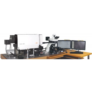
3D Scanning Laser Confocal Raman Microscope Confotec™ NR500
Features
spectral resolution up to 0.006 nm Use of inverted and upright microscopes is possible. Telescope with variable magnification for adapting laser beams to entrance pupils of microobjectives from 3 to 12 mm. Polarized measurements. High sensitivity at low power of laser excitation (from ?W to mW). Reflection module for simultaneous obtaining of 3D image in reflected light. Transmission measurements option. Fully automated control of the system. High temporal and temperature stability is provided by modular rigid and rod design. No fiber optics that decrease some optical parameters (transmission, wave front, polarization). Ring illumination for combination with AFM. |
Signal decrease from 90 % to 10 % at 200 nm, λ=514 nm, 100Х immersion lens |
Spatial resolutions?
|
|||
Laser?
|
Objective lens: magnification and numerical aperture NA |
XY Spatial resolution?
|
Z Spatial resolution?
|
488 nm |
100X, NA = 0.95?
|
295 nm |
450 nm |
633 nm |
100X, NA = 0.95?
|
395 nm |
590 nm |
785 nm |
100X, NA = 0.95?
|
560 nm |
750 nm |
Spectral range for Raman spectra detection: |
30 cm -1 ~ 6000 cm -1 (depends on the wavelength of the excitation laser)
|
Spectral resolution: |
0,25 cm-1 (75 l/mm Echelle grating)?
|
Sensitivity: |
peak of the 4th order of the Raman spectrum of Si is detected within 1 min |
Modes of scanning: |
- Fast mapping: scanning of a laser beam along the surface of a sample with XY galvanoscanner
- shifting a sample with XY motorized scanning stage near a fixed laser beam
- combined mode for fast obtaining panoramic images with high spatial resolution: XY scanner (Fast mapping) + motorized microscope stage |
Maximum scanning field size in the "Fast mapping" mode: |
XY 150 х 150 μm (with objective lens 100х)?
|
Period of registration for one frame 150 х 150 μм in the "Fast mapping" mode: |
3 s. / 1001 х 1001 pixels |
PC control: |
fully automated |
Microscope |
|
*Type, model: |
inverted Nikon Ti-S and upright Nikon Ni-U |
Motorized scanning stage: |
automated |
- travel range |
114 х 75 mm |
- accuracy (1 mm of shift) |
0.06 μm |
- XY repeatability |
± 1 μm |
- minimal step |
0.02 μm |
Micro objective lens: |
100х NA-0.95 40х NA-0.7520х NA-0.50 and other |
Z-scanner: |
piezo scanner |
- objective translation range |
80 μm |
- objective translation step |
50 nm |
- repeatability | < 6 nm
|
High-resolution digital video camera: |
digital color CCD camera |
- sensor |
1/2", 2048 x 1536 pixels |
- ADC |
10 bit, speed 12 frames/s |
Laser radiation delivery: |
three-position turret |
* other types of inverted or upright microscopes available | |
Optical-mechanical unit (OMU) |
|
Optimized optics for spectral range: |
325 - 1050 nm (UV-VIS-NIR) 400 - 1100 nm (VIS-NIR) |
Laser radiation delivery: |
triple- and quintuple input port |
Laser beam attenuator: |
automated unit with VND filter, 0 - 3D |
Polarizers (excitation channel) and analyzer (detection channel): |
Glan-Taylor prism (automated unit) |
Laser beam expander: |
automated vario telescope, magnification factor 1.0 ? 4.0x?
|
Half-wave (λ/2) plate positioner: |
automated three- / five-postion |
Raman filter positioner: |
automated three- / five-postion |
Interference filter positioner: |
automated six-position |
Pre-pinhole objective lens positioner: |
automated three-coordinated (X, Y, Z) |
Image Monochromator-Spectrograph MS5004i |
|
Optical configuration: |
vertical |
Focal length: |
520 mm |
Magnification: |
1.0 vertical, 1.0 horizontal |
Vertical spatial resolution: |
< 20 μm |
Flat field size: |
28 х 5 mm |
Stray light: |
1 х 10-5 (20 nm from laser line 633 nm)?
|
Diffraction grating unit: |
automated four-position turret |
Spectral resolution: (wavelength 500 nm, CCD pixel 12 x 12 μm) |
0.25 cm-1 (Echelle grating 75 l/mm) 0.9 cm-1 (grating 1800 l/mm) |
Spectral slits: |
automated confocal pinhole, smoothly regulated from 0 to 1.5 mm |
- entrance |
automated, smoothly regulated from 0 to 2 mm |
- output |
automated, smoothly regulated from 0 to 2 mm |
Ports: |
1 input, 2 output |
Output ports switching: |
automated output mirror |
Spectral camera for spectrograph
|
|
Type: |
digital CCD camera |
Sensor type: |
back-thinned CCD sensor 2048 х 122 pixels |
Pixel size: |
12 x 12 μm |
Pixel area size: |
24.576 x 1.464 mm (width x height) |
Spectral response range: |
from 200 to 1100 nm |
Cooling with temperature stabilization: |
two-stage Peltier element, min ? 45 °С
|
ADC: |
16 bit |
Sensivity: |
1 photon for 1 ADC reading (at the max. sensivity of 650 nm) |
Dynamic range: |
not less than 10 000 |
Fast scanning unit X, Y
|
|
Scanners: |
galvanometer scanners with X, Y mirrors |
Scanning modes: |
raster high-speed and start-stop |
Positioning accuracy: |
30 nm |
Scanning area: |
150 μm х 150 μm (with 100Х objective lens)?
|
Scanning speed: |
3 s/frame 1001 х 1001 pixels |
Unit of confocal laser microscope
|
|
Pre-pinhole objective lens positioner: |
automated three-coordinate (X, Y, Z) |
Confocal pinhole: |
automated confocal pinhole, smoothly regulated from 0 to 1.5 mm |
Detector: |
Hamamatsu Photosensor module H6780-01? |
Lasers |
||
Type of laser: |
Up to 5 lasers can be used simultaneously |
|
|
Wavelength, nm?
|
Output power, mW?
|
He-Cd Laser (Single Mode (TEM00) He-Cd): |
325 |
15,30,40,50 |
Diode-pumped Laser (DPSS): |
473 |
25,50 |
Diode-pumped Laser (DPSS): |
532 |
25,50 |
He-Ne Laser: |
633 |
10 |
Diode-pumped laser (DPSS): |
785 |
80 |
|
Using of other types of lasers with wavelength from 350 to 850 nm is possible |
|

| 품명 | NR500 |
|---|---|
| 모델명 | NR500 |
| 법에 의한 인증·허가 등을 받았음을 확인할 수 있는 경우 그에 대한 사항 | 상품페이지 참고 |
| 제조국 또는 원산지 | Belarus |
| 제조자 | SOL instruments Ltd. |
| A/S 책임자와 전화번호 또는 소비자상담 관련 전화번호 | 02-355-5963 |
사용후기가 없습니다.
상품문의가 없습니다.
등록된 상품이 없습니다.


